
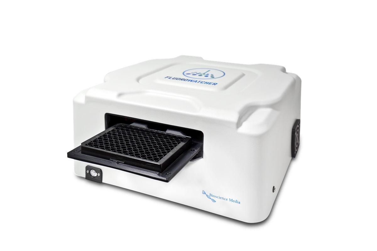
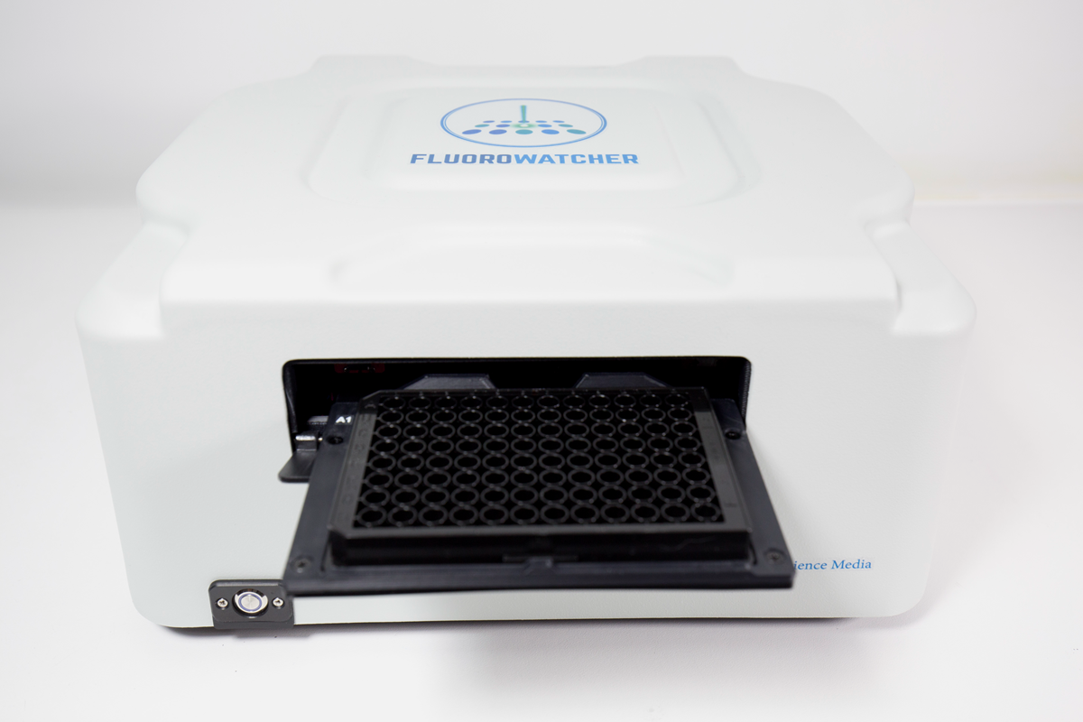
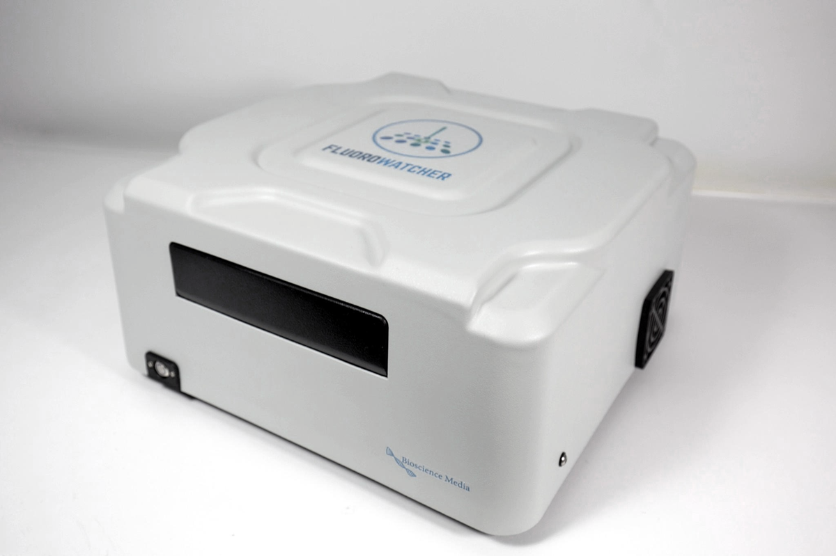

Fluorowatcher takes fluorescence images of microarrays and measures the spot intensities with the QuantoPic software. The user interprets the results.
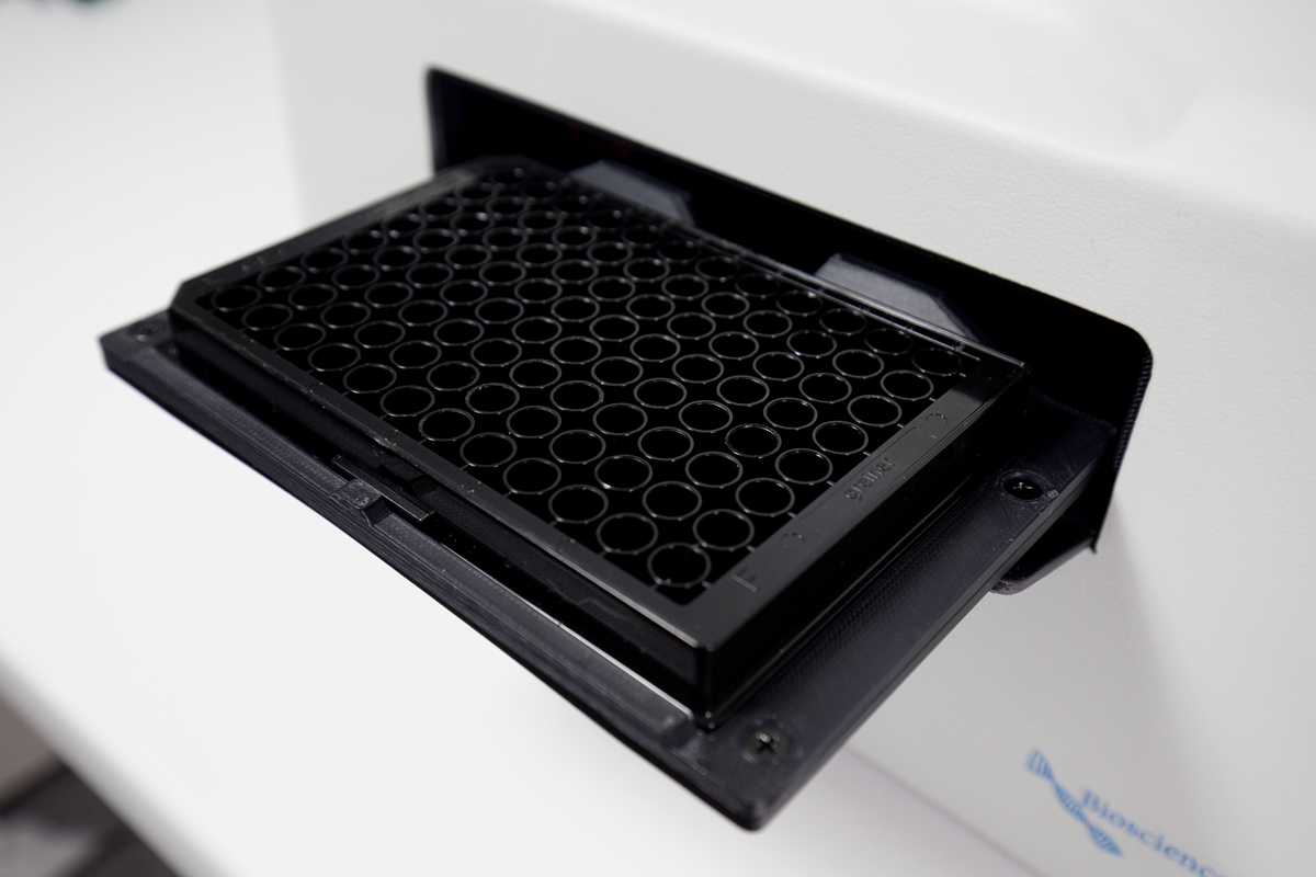
It works with two fluorescence channels, Cy3 and Cy5, and reads 96-well plates, 12×8 well strips and microscope slides (with an adapter). Imaging is from below, so the plates need a transparent bottom.
A high-sensitivity CMOS camera gives a high signal-to-noise ratio at short exposure times. A built-in emission filter separates the fluorescence. An automated mechanism ejects the plate.
Fluorowatcher is compact and fits into an airline carry-on bag.
Fluorowatcher is designed and manufactured under a quality management system based on ISO 13485.
- Two fluorescence channels: Cy3 and Cy5
- Laser light source, lifetime over 10 000 hours
- 3 MP CMOS camera, 6 µm per pixel, field of view 7.7 × 7.7 mm
- 96-well plates, 12×8 well strips, up to 4 microscope slides
- Automated plate ejection
- Software-controlled focus; programmable exposure up to about 30 s
- Control of exposure, XYZ stage and channel switching from QuantoPic
- Enclosed laser with a two-channel lid interlock that works independently of the firmware
- QuantoPic is included. It controls the instrument and analyses the images.
- AI Array Finder: automatic microarray grid detection and alignment
- Grid layout and region mapping
- Automated image acquisition and positioning
- Spot intensity (total, mean, median) with background correction
- Qualitative and quantitative assays, including 4PL and 5PL calibration curves
- Point-and-shoot imaging at specified locations
- Export of results to PDF, CSV and Excel
- Product brochure (PDF)
- User manual and conformity documentation for your instrument: please contact info@biosciencemedia.lv and include the model and serial number, if available.
| Imaging mode | Fluorescence, 2 channels: Cy3 and Cy5 |
| Light source | Diode laser, green and red |
| Light source lifetime | Over 10 000 hours |
| Measurement range | 0 to 65 535 (arbitrary units) |
| Vessels | 96-well plate, 12×8 well strip, 4 microscope slides (with an adapter) |
| Camera | 3 MP, CMOS |
| Standard image size | 1280 × 1280 pixels |
| Resolution | 6 µm per pixel |
| Field of view (standard image) | 7.7 × 7.7 mm |
| Focus | Manual (software-controlled) |
| Exposure | Up to about 30 s (programmable) |
| Image formats | PNG or TIFF, 16-bit |
| Data interfaces | USB 2.0 (instrument control), USB 3.0 (camera) |
| Software | QuantoPic, included |
| Size (W × D × H) | 330 × 345 × 170 mm |
| Weight (without power supply) | Up to 8 kg |
| Power supply | External power supply XP Power AKM90PS24 (CE marked): input 100–240 V AC, 50/60 Hz; output 24 V DC, 3.75 A, 90 W. Instrument input: 20–24 V DC. |
| CPU | Intel Core i7 with AVX2 support |
| RAM | 8 GB |
| Graphics card | Nvidia GeForce GTX 1050 Ti 4 GB or better (Nvidia GeForce 10, 16, 20 or 30 series only) |
| Storage | SSD, 256 GB |
| Operating system | Windows 11, 64-bit |
- Research use only (RUO). Not a medical device and not an in vitro diagnostic medical device.
- CE marking: EMC Directive 2014/30/EU, Machinery Directive 2006/42/EC, RoHS Directive 2011/65/EU.
- Designed and manufactured in Riga, Latvia, by SIA Bioscience Media.